ヨハンセンは、ドイツ読みで、英語でjohnson法(ジョンソン法)と呼ばれています。
FEMORAL NECK
Mediolateral projection
Johnson position
This method of obtaining an axiolateral view of the femoral neck is employed when the opposite femur cannot be adjusted for one of the two preceding positions.
Film: 8 '' X 10'' placed lengthwise in a vertical position.
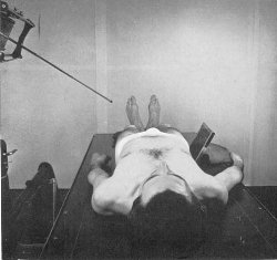
Position of part
When necessary, elevate the pelvis on folded sheets or a firm pillow so as to place the greater trochanters about 6 inches above the tabletop (2 inches above the center line of the film). This is to allow for the laterodorsal angle of the central ray. Support the extremities at hip level on pillows or sandbags. Adjust the body in a true antero-posterior position. Place the cassette in the vertical position along the lateral surface of the hip and center the transverse line of the cassette to the greater trochanter, which should be approximately 2 inches above the longitudinal line. Tilt the cassette backward 25 degrees from the vertical and support it in position with sandbags or other suitable support.
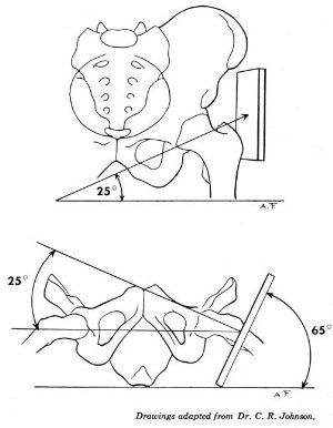
Central ray
The central ray is directed to the greater trochanter at a double angle 25 degrees toward the head plus 25 degrees posteriorly. It enters at about the midsagittal plane of the thigh.
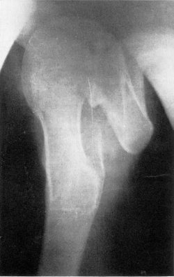
Structures shown
An axiolateral view of the head, neck, and trochanteric region of the femur.
Johnson, C.R.: A new method tor roentgenographic examination of the upper end of the femur, J.
Bone Joint Surg. 30:859-866, 1932.